What Are X-Ray Scintillators?

What Are X-Ray Scintillators?

If you work with X-ray imaging equipment, you have probably come across the term before; but what are x-ray scintillators, exactly? They are materials that respond to X-ray radiation and re-emit that energy as visible photons, making them a vital component in a wide variety of X-ray imaging devices. Put simply, they convert invisible radiation into visible light that imaging systems can then detect and record. Used across both medical and security applications, these materials sit at the core of modern X-ray imaging technology and the quality of the scintillator has a direct bearing on the quality of the image produced.

Why a Poor Quality Scintillator Can Compromise Your Imaging Results

If the scintillator at the heart of your imaging device is inconsistently manufactured or incorrectly specified, the consequences ripple through everything that follows. Uneven coating, impure raw materials, or a poorly controlled production environment can all lead to variable light output, inconsistent afterglow behaviour, and ultimately unreliable images. For organisations working in medical or security imaging, where precision and consistency are non-negotiable. This is a risk that cannot be overlooked. The quality of the scintillator is not a minor detail; it defines the integrity of the entire imaging process.

How X-Ray Scintillators Work

The process of scintillation begins the moment X-ray energy strikes the phosphor material within the scintillator. That energy is absorbed and then re-emitted as visible photons – light that can be captured and processed by the imaging system downstream.

The phosphor compounds used in scintillators are often rare-earth materials, and each is designated by the letter P followed by a number, which identifies its specific characteristics and performance profile. The behaviour of any given phosphor, including how brightly it emits, at what wavelength, and for how long, is shaped in part by the addition of an activator compound.

Activators play a particularly important role in controlling afterglow, which is the continued emission of light after the X-ray source has been removed. Depending on the application, an activator can be used to prolong afterglow where that is desirable, or to quench and shorten it where a faster response is required. Getting this balance right for the specific application is one of the key considerations in selecting the correct scintillator.

The Different Types of X-Ray Scintillator

Analytical Components manufactures three core product types, each serving a distinct imaging requirement.

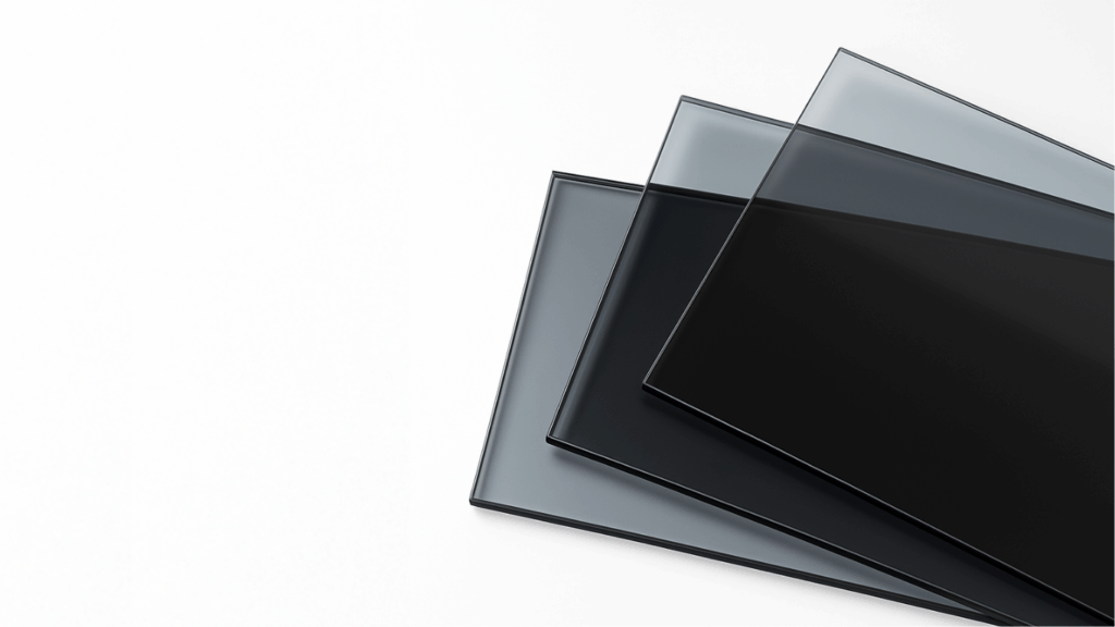
  • X-Ray Phosphor Scintillators are the primary product, designed to respond directly to X-ray energy and re-emit it as visible light. They can be incorporated into a wide variety of X-ray imaging devices across medical and security sectors.
  • Phosphor Screen Scintillators follow the standard phosphor designation system; the letter P followed by a number, with different phosphor types offering different emission and afterglow characteristics to suit the requirements of different imaging systems. Each phosphor is often a rare-earth compound, with the activator playing a key role in tuning performance.
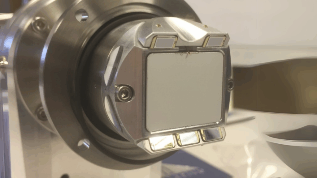
  • EBSD Phosphor Screens are a specialist product used within scanning electron microscopes (SEM). Electron backscatter diffraction (EBSD) is a technique used to collect and explore quantitative microstructure analysis data, including crystal orientation, phase grain statistics, phase and strain conditions, and defects. The phosphor screen is central to capturing the diffraction patterns that make this level of analysis possible.

What Are X-Ray Scintillators and Why Does Manufacturing Quality Matter So Much?

The answer to what x-ray scintillators are is relatively straightforward, but understanding why manufacturing quality matters is equally important. Producing scintillators to a consistently high standard requires both the right environment and the right processes.

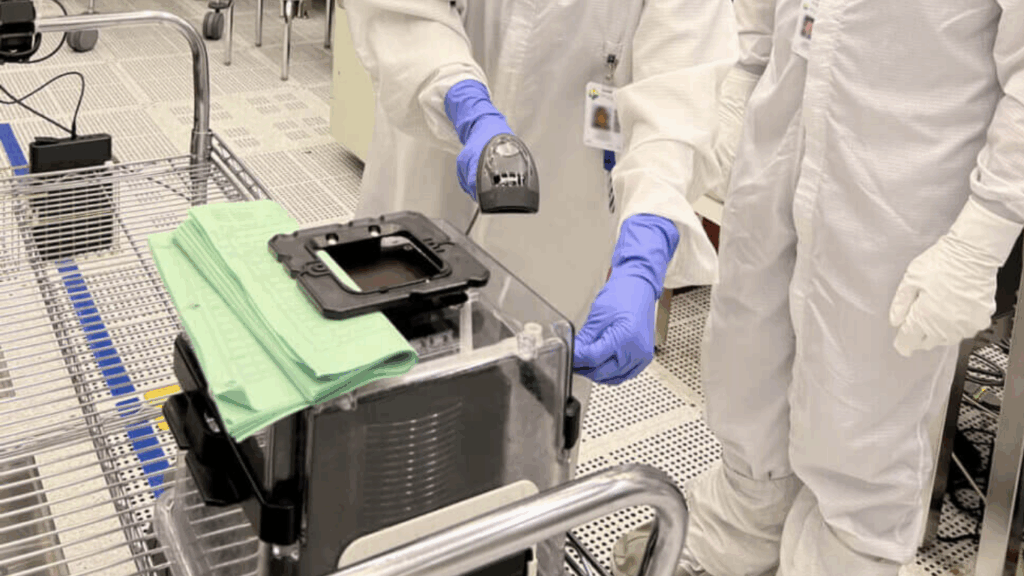
Analytical Components manufactures its products within a Class 10,000 (ISO 7) cleanroom. This controlled environment minimises contamination during the coating process and ensures that the raw materials used meet the high standards needed for uniform, consistent output. The coating process itself demands specialist equipment and highly trained staff, both of which are central to delivering products that perform reliably whether they form part of a large production run or a bespoke one-off piece.

The company holds ISO 9001:2015 accreditation, which means it has formally demonstrated the ability to consistently provide products and services that meet both customer and regulatory requirements. Clients are also encouraged to visit the production facility directly, and frequent updates are available via telephone or email throughout the manufacturing process. Where required, non-disclosure agreements are offered as standard, reflecting a genuine respect for customer confidentiality.

For anyone sourcing scintillation components, understanding what x-ray scintillators are is only the starting point. The manufacturing process, the cleanroom environment, and the quality controls surrounding production are what ultimately determine whether the scintillator in your imaging device will perform as it should, consistently, reliably, and to specification.

Frequently Asked Questions

What Are X-Ray Scintillators?

X-ray scintillators are materials that absorb X-ray radiation and re-emit that energy as visible photons, making them an essential component in medical and security imaging devices.

What do x-ray scintillators do?

They absorb X-ray radiation and re-emit that energy as visible photons, allowing imaging systems to capture and process the resulting light signal.

What materials are x-ray scintillators made from?

They are typically made from rare-earth phosphor compounds. An activator is often added to the phosphor to influence its emission behaviour, including the duration of afterglow.

What is afterglow and why does it matter?

Afterglow is the continued emission of light from a scintillator after the X-ray exposure has ended. Depending on the application, this can be a useful property or an unwanted one. Activator compounds within the phosphor can be used to either prolong or shorten afterglow accordingly.

What is the phosphor designation system?

Phosphors are designated by the letter P followed by a number. This system identifies the specific phosphor type and its associated performance characteristics, allowing engineers and buyers to select the correct material for their imaging application.

What is an EBSD phosphor screen used for?

An EBSD phosphor screen is used within a scanning electron microscope to support electron backscatter diffraction analysis. This technique collects quantitative microstructure data including crystal orientation, phase grain statistics, strain conditions, and defects.

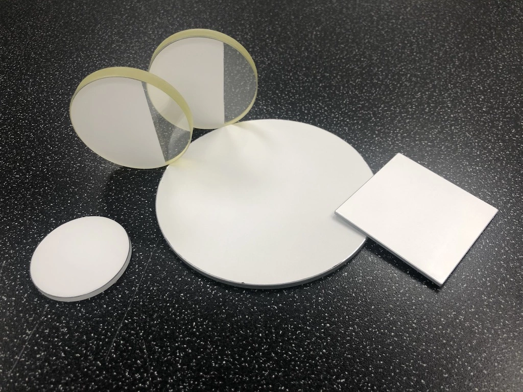
Can scintillators be manufactured to a bespoke specification?

Yes. Specialist manufacturers are able to produce scintillation products ranging from large-scale production orders to bespoke one-off pieces, each manufactured to the individual customer’s requirements.

Understanding Afterglow in Scintillator Materials

Understanding afterglow in scintillator materials is essential for improving the accuracy and efficiency of imaging and detection systems. Afterglow, also known as persistence, refers to the continued emission of light after the excitation source has been removed. While this lingering light may seem harmless, it can interfere with data clarity, image resolution and timing precision. At Analytical Components, we take a deep interest in how afterglow affects performance and how it can be minimised through careful material design and manufacturing control.

A close-up photo of a clear glass square on a smooth surface with soft lighting.

What Is Afterglow in Scintillator Materials

When radiation or energetic particles strike a scintillator, electrons in the phosphor become excited to higher energy levels. Ideally, they should return to their ground state immediately, releasing energy in the form of visible light. However, in practice, some electrons become trapped in defects or imperfections within the material. These trapped electrons are released slowly, continuing to emit light after the excitation has stopped.

This delayed emission is what we describe as afterglow. Understanding afterglow in scintillator materials helps manufacturers and scientists refine both the chemical composition and structural purity of the phosphor layer to create faster and more reliable responses.

Why Afterglow Matters

When it comes to imaging and detection systems, particularly those requiring high speed or high resolution, afterglow can significantly impact performance. In X-ray imaging, for instance, it can cause residual light from one frame to carry over into the next, leading to blurred or ghosted images. In electron microscopy and other analytical techniques, persistent light can obscure fine structural details or distort readings.

Reducing afterglow ensures that each signal corresponds precisely to its intended event. It prevents one exposure from influencing the next, preserving image clarity and measurement integrity. At Analytical Components, our phosphor screen coatings are engineered to provide the perfect balance between brightness, decay time and minimal afterglow, ensuring dependable performance in demanding imaging environments.

Factors That Influence Afterglow

The level of afterglow in a scintillator material depends on several interconnected factors. The composition of the phosphor determines its luminescent efficiency and decay behaviour. For example, materials such as P43 phosphor offer excellent brightness but moderate persistence, while P47 phosphor provides a faster decay and reduced afterglow.

Purity is another key factor. Even trace impurities or structural defects within a crystal lattice can act as traps for electrons, extending decay time. Temperature also affects behaviour, as higher temperatures can help trapped electrons escape more quickly, reducing persistence. Finally, coating thickness and uniformity influence optical consistency. A precisely applied phosphor coating ensures predictable performance across the entire screen surface.

Every one of these parameters is carefully managed at Analytical Components. Our cleanroom-controlled coating processes ensure the highest possible uniformity and material stability, allowing us to deliver products that perform consistently across all imaging conditions.

Measuring and Characterising Afterglow

Evaluating afterglow involves measuring the light that continues to be emitted after the excitation source is removed. This data is often displayed as a decay curve, showing how quickly the light intensity drops over time. By comparing these curves, manufacturers can determine which materials are best suited to specific imaging requirements.

Low-afterglow scintillators are critical for applications that demand rapid response times, while others may prioritise light output or durability. At Analytical Components, we use this data-driven understanding to guide our material choices and coating methods, ensuring that each product meets the precise requirements of its intended use.

Achieving the Right Balance

Minimising afterglow entirely is rarely possible without affecting other performance aspects such as brightness or decay rate. The art of scintillator design lies in achieving the optimal compromise between light yield and response speed. Bright phosphors often have longer decay times, while faster materials can produce slightly lower light output.

Through continuous testing and refinement, we have developed methods to control these trade-offs effectively. Our coatings maintain exceptional brightness while keeping afterglow within tightly controlled limits. This careful balance allows scientists and engineers to work with materials that deliver both clarity and consistency, even under high-speed imaging conditions.

Applications Where Afterglow Control Matters

Understanding afterglow in scintillator materials is particularly important in high-precision imaging systems. In X-ray inspection and non-destructive testing, it ensures clear images of moving parts or materials. When performing electron microscopy, it allows accurate data capture from dynamic samples. In medical imaging and security screening, low-afterglow materials improve image refresh rates and diagnostic accuracy.

These benefits translate directly to the reliability of results and the quality of analysis. Every scintillator produced by Analytical Components is designed with these applications in mind, offering dependable performance across scientific, industrial and research environments.

The Analytical Components Approach

At Analytical Components, we combine technical expertise with a commitment to quality manufacturing. Every scintillator and phosphor screen we produce is made in an ISO 9001 certified cleanroom environment. This ensures that coatings are applied with maximum precision and that every product achieves the same high standard of optical clarity and uniformity.

Our ongoing research into material performance, coating methods and decay characteristics allows us to stay at the forefront of scintillator technology. Whether you require a fast-response phosphor for electron imaging or a high-efficiency screen for X-ray detection, we can develop a solution that delivers the performance your work demands.

Image of a Scintillator

Get In Touch

Understanding afterglow in scintillator materials is central to improving image clarity, measurement precision and detector performance. It reflects the delicate balance between material science, coating technology and optical engineering. At Analytical Components, we focus on controlling every factor that influences afterglow, from material purity to coating uniformity, ensuring our products meet the highest expectations for speed and quality.

At Analytical Components, we don’t just supply high-performance scintillators. We back that up with technical insight, cleanroom manufacturing, and responsive customer support. If you ever have questions about performance, replacement, or handling, we’re only a message away. You can learn more about what we do on our about us, or reach out directly to speak to someone on the team.

Get in touch today via our contact form, email us at info@analyticalcomponents.uk, or call us at +44 (0) 1424 850004 to discuss your requirements.

How Reflective and ITO Coatings Enhance Light Output

Understanding how reflective and ITO coatings enhance light output is essential to producing high-performance scintillator and phosphor screens. These coatings define the brightness, clarity and efficiency of modern imaging systems. It plays a central role in how light is captured, redirected and transmitted to the detector. At Analytical Components, our precision manufacturing processes and ISO-certified cleanroom environment ensure that both reflective and ITO coatings deliver exceptional optical and electrical performance for a wide range of scientific applications.

Reflective and ITO Coatings

Understanding Reflective and ITO Coatings

Reflective and ITO, or Indium Tin Oxide, coatings each serve a distinct function in the structure of a phosphor screen. A reflective layer, often made from evaporated aluminium, is designed to redirect light. This would otherwise be lost through the rear of the phosphor. This helps maximise signal strength and brightness.

An ITO coating, by contrast, is a conductive but transparent layer. It allows charge to dissipate without blocking visible light. This property makes it essential for electron-based imaging systems such as Electron Backscatter Diffraction and Transmission Electron Microscopy.

Through careful control of coating thickness and surface preparation, Analytical Components achieves a balance between conductivity, reflectivity and optical precision that enhances imaging performance across every product line.

How Reflective Coatings Improve Brightness

Reflective coatings play a key role in boosting the apparent brightness of a phosphor screen. When incoming radiation or electrons excite the phosphor, light is emitted in all directions. Without a reflective layer, a large portion of that light escapes backwards, reducing output efficiency. Applying a thin, uniform aluminium coating redirects this light forward, increasing signal intensity without the need for thicker phosphor layers.

At Analytical Components, aluminium coatings are applied in a cleanroom to achieve extremely high reflectivity and consistency. Each coating is carefully monitored to maintain optical uniformity and prevent scattering. This precision ensures bright, stable images and reliable calibration over extended operational use.

The Advantages of ITO Coatings in Electron Imaging

While reflective coatings increase optical brightness, ITO coatings are essential for maintaining image stability in electron-based systems. In environments where electron beams interact with phosphor surfaces, static charge can accumulate, distorting the image or reducing clarity.

An ITO layer provides an effective way to dissipate that charge while allowing light to pass freely through to the detector. Its combination of transparency and conductivity makes it ideal for sensitive scientific instruments. At Analytical Components, our vacuum deposition techniques produce uniform, finely controlled ITO layers that ensure electrical performance without compromising optical quality. This meticulous attention to coating integrity is a hallmark of our manufacturing process.

Balancing Coating Thickness and Optical Performance

Optimising how reflective and ITO coatings enhance light output depends on precise control of coating thickness. If a reflective layer is too thick, it can scatter light and reduce sharpness. If an ITO layer is too thin, it may not conduct efficiently across the surface.

Our manufacturing process allows coating parameters to be tailored for each application. X-ray scintillator screens may require thicker reflective coatings to achieve maximum brightness, while electron imaging screens benefit from thinner, highly transparent ITO layers. Every product is tested for optical consistency, adhesion and performance before leaving our facility, ensuring reliability from first use.

Applications Across Imaging and Detection

Reflective and ITO coatings are integral to performance across a wide spectrum of imaging technologies. When X-ray imaging, reflective coatings increase light yield and enhance contrast. In electron microscopy, ITO coatings prevent charging effects that can interfere with precision imaging. In medical, industrial and scientific detectors, both coating types work together to maximise light capture and maintain consistent results.

By continuously refining how reflective and ITO coatings enhance light output, Analytical Components supports the development of instruments that deliver higher accuracy, sharper resolution and improved efficiency.

Cleanroom Manufacturing for Maximum Precision

The environment in which coatings are applied is just as critical as the coating materials themselves. Contamination can lead to scattering points or uneven surfaces that reduce optical efficiency. To avoid these issues, all coatings at Analytical Components are applied in an ISO 9001 certified cleanroom environment.

This level of precision ensures the highest possible quality in both reflective and conductive coatings, with consistent results across every production batch. Our cleanroom procedures, advanced vacuum systems and rigorous quality checks enable us to meet the demanding standards expected by leading manufacturers and research institutions.

Continuous Development in Coating Technology

The science behind how reflective and ITO coatings enhance light output continues to evolve alongside advances in detector and imaging technology. At Analytical Components, we are committed to ongoing research and development. We are always exploring new coating techniques and materials to achieve superior performance. Our ability to work with a range of substrates, from glass and fibre optic to silicon and metal, allows us to deliver tailored solutions for every imaging challenge.

Whether you need high-reflectivity aluminium coatings for X-ray detection or finely tuned ITO coatings for electron imaging systems, we can provide the expertise and precision required for outstanding optical performance.

Reflective and ITO Coatings

Conclusion

Producing coatings that enhance light output demands precision, expertise and an understanding of how each layer interacts with light and charge. Reflective and ITO coatings are not simply surface treatments. They are integral to the performance of the entire imaging system, from X-ray detection to electron microscopy.

At Analytical Components, we combine deep technical knowledge with cleanroom manufacturing and responsive customer support. Our coatings are developed to the highest standard, ensuring every scintillator or phosphor screen we produce delivers exceptional optical and electrical performance. If you would like to discuss how our reflective or ITO coatings can support your next project, we’re always ready to help.

Get in touch today via our contact form, email us at info@analyticalcomponents.uk, or call us at +44 (0) 1424 850004 to discuss your requirements.

X Ray Scintillators in the UK

X ray scintillators are a critical part of modern imaging systems. Whether used in research labs, industrial inspection equipment, medical diagnostics or security screening, these materials allow high energy radiation to be visualised with precision and clarity. In the UK, the demand for reliable, high performance scintillator solutions continues to grow across a wide range of sectors. At Analytical Components UK, we support this need by supplying custom coated scintillator screens designed for technical accuracy, consistency and durability.

X Ray Scintillators in the UK

What Are X Ray Scintillators?

A scintillator is a material that emits light when it is exposed to radiation. In x ray systems, scintillators absorb incoming x ray photons and re emit them as visible light. This light can then be captured by a sensor or camera, producing a detailed image for analysis. The quality of that image depends heavily on the performance of the scintillator screen itself.

Different applications require different types of scintillators. For example, electron microscopy setups often rely on phosphor screens optimised for electron backscatter diffraction, while high speed medical imaging may use materials with fast response times and minimal afterglow. The choice of scintillator must be guided by the intended use, the resolution requirements of the system and the conditions under which it operates.

Why They Are Important in UK Industries

Across the UK, x ray scintillators are in use every day by scientists, engineers and technicians. In manufacturing, they help identify microstructural defects in materials before they reach the final product. In research laboratories, they enable precise crystal orientation mapping through EBSD techniques. In hospitals and clinics, they help clinicians make fast and accurate diagnoses from high quality x ray scans. In airports and transport hubs, they support security teams by revealing concealed threats with clarity and speed.

This widespread use calls for scintillator screens that are not only effective, but also consistent, durable and easy to integrate. UK customers increasingly value local technical support and fast delivery. This is as well as tailored solutions that fit their specific detection systems.

Our Role at Analytical Components

We specialise in providing high quality x ray scintillator screens for commercial and scientific use. Each screen is manufactured to precise standards using cleanroom coating processes to ensure maximum light output, surface uniformity and long term reliability. From phosphor coated substrates to EBSD specific screens, our products are designed with both performance and practicality in mind.

What makes our approach different is our technical background and hands on experience with a wide range of detection systems. We do not offer generic off the shelf products. Instead, we work closely with clients to understand the exact requirements of their setup and supply solutions that integrate seamlessly with existing workflows.

Being based in the UK also allows us to offer quick turnaround. We give direct communication and quality control you can rely on.

X Ray Scintillators in the UK

Applications in Detail

Many of our clients are involved in materials characterisation, using techniques such as SEM and EBSD to study the properties of metals, ceramics, semiconductors and other materials. In these cases, our phosphor screens provide high brightness and stability, helping researchers gather detailed structural data with confidence.

Others operate in non destructive testing, where our scintillator screens are used in flat panel x ray detectors to inspect parts for internal defects without damaging them. We also support developers of medical and veterinary imaging devices. We provide phosphor coatings that enhance image contrast and reduce exposure times for patients and animals alike.

What to Consider When Selecting a Scintillator

The key factors to consider include the type of radiation involved, the required image resolution, the operating environment and the type of sensor being used. Scintillator thickness, coating material, substrate type and mounting method can all affect performance.

This is where our team can offer guidance. With our technical knowledge and manufacturing capabilities, we can recommend the best options for your specific goals and constraints.

X Ray Scintillators in the UK

Contact Analytical Components for Expert Scintillation Solutions

X ray scintillators may not always be visible in the final product or research paper, but their role is foundational. Clearer images mean better decisions, whether in a clinical setting, a laboratory or a factory. At Analytical Components UK, we are proud to support this progress by offering scintillator solutions that meet the highest standards of performance and reliability.

At Analytical Components, we are committed to delivering high-quality scintillation solutions tailored to your specific industry requirements. Whether you need X-ray scintillators, EBSD phosphor screens, or custom phosphor-based detection systems, our expert team is ready to assist you. With our ISO 9001:2015 accredited processes and state-of-the-art cleanroom facilities, you can trust us to provide precision-engineered scintillation components.

Need expert advice? Get in touch today via our contact form, email us at info@analyticalcomponents.uk, or call us at +44 (0) 1424 850004 to discuss your project requirements. Whether you require phosphor screens for microscopy or X-ray scintillators for imaging applications, we can help.

Enhance your imaging capabilities with precision-engineered scintillators from Analytical Components.

Comparing P43 and P47 Phosphors for Imaging Applications

Comparing P43 and P47 phosphors for imaging applications is vital for researchers, laboratories and manufacturers who rely on precision results. The choice between these two phosphor types can determine whether an imaging system delivers high sensitivity, rapid frame rates, or consistent clarity across repeated tests. At Analytical Components, we provide expertly coated phosphor screens that are designed to meet the demanding requirements of advanced imaging, ensuring that every screen we supply delivers both reliability and performance.

Why Phosphor Choice Matters

Phosphor screens play a central role in scintillation. When X rays or electrons strike the screen, the phosphor layer emits visible light that can be captured by cameras or detectors. The properties of the phosphor directly influence how much light is emitted, how long that light persists, and the colour of the emission. This means the choice of phosphor is not a minor detail but one of the most important decisions when specifying a scintillator screen. At Analytical Components, our expertise lies in providing the right phosphor for the right application, supported by our ISO 9001 certified cleanroom manufacturing.

Understanding P43 and P47

When comparing P43 and P47 phosphors for imaging applications, the most noticeable difference lies in their decay times and light output.

  • P43 phosphor emits a green light at around 545 nanometres and is known for its strong brightness and efficiency. It provides an excellent signal for detectors but has a relatively longer decay time, typically around one millisecond. This makes P43 highly effective for imaging setups that prioritise light yield and can tolerate slightly slower response times.
  • P47 phosphor emits in the blue white region at around 400 nanometres. While its brightness is lower than P43, it has an exceptionally fast decay time, often in the range of tens of nanoseconds. This property makes P47 an outstanding choice for high speed imaging, where avoiding afterglow and preventing overlapping signals are essential.

At Analytical Components, we offer both P43 and P47 phosphors applied with precision coating techniques to ensure uniformity and durability, giving you confidence in the performance of your imaging system.

Decay Time and Imaging Speed

For many users, the deciding factor in comparing P43 and P47 phosphors for imaging applications is decay time. In fast imaging environments, even a small amount of persistence can cause afterimages or ghosting that compromise results. This is why P47 is so often chosen for experiments such as Particle Imaging Velocimetry or time resolved microscopy. On the other hand, if sensitivity is more important than speed, P43 provides higher efficiency, producing a stronger and brighter signal that makes detection easier in low light conditions.

At Analytical Components, we help clients analyse the trade offs between brightness and speed. Our team works closely with research groups, medical imaging specialists and industrial partners to determine which phosphor delivers the most accurate results for their systems.

Spectral Output and Detector Sensitivity

The emission colour of each phosphor also matters. Detectors often have peak sensitivities at particular wavelengths. P43, with its green emission, is very well suited to many CCD and CMOS detectors, which often have high sensitivity in the green region of the spectrum. P47, with its blue emission, may require detectors that perform better at shorter wavelengths. This means that the decision is not just about decay time but also about how the phosphor interacts with the rest of the imaging system.

Our phosphor screen scintillators at Analytical Components can be manufactured with additional coatings, such as Indium Tin Oxide or reflective aluminium, to enhance performance and ensure that the maximum amount of emitted light is directed towards the detector. This level of customisation ensures that whether you select P43 or P47, the screen you receive will perform exactly as you need it to.

Matching the Phosphor to the Application

When comparing P43 and P47 phosphors for imaging applications, the final choice often comes down to the context in which the screen will be used.

  • P43 is the better choice for applications that require longer exposure times, higher sensitivity and brighter images. It is commonly used in systems where image quality and clarity matter more than frame rate, such as certain forms of X ray imaging or slower scanning electron microscopy setups.
  • P47 is the phosphor of choice for high speed imaging and time resolved studies. Its ultra fast decay ensures that each frame is clean, without interference from the previous exposure. This makes it indispensable in applications like fast electron microscopy or high frame rate optical imaging.

At Analytical Components, we understand that no two applications are identical. That is why we offer both phosphors across a range of substrates including glass, fibre optic plates, silicon and metal, ensuring you can build a scintillator screen that is perfectly suited to your needs.

The Importance of Cleanroom Manufacturing

Another critical factor in performance is the quality of the coating itself. Even the best phosphor can fail if it is not applied with consistency and precision. At Analytical Components, we operate from a Class 10,000 ISO 7 cleanroom, accredited to ISO 9001:2015 standards. This environment ensures that every screen we produce has a uniform layer of phosphor, free from contamination or inconsistencies. For our clients, this means dependable, repeatable results and confidence that their imaging equipment will perform at its best.

Supporting Innovation and Custom Development

We also recognise that many clients require bespoke solutions. Some projects may demand specific coating thicknesses, additional conductive layers or unusual substrate sizes. At Analytical Components, we specialise in providing both one off prototypes and larger production runs. By working closely with your research or engineering team, we ensure that whether you need a single screen for testing or a full batch for integration, your requirements are fully met.

Get In Touch

Comparing P43 and P47 phosphors for imaging applications is about more than simply choosing between green and blue light. It is a decision that influences speed, brightness, clarity and the overall performance of your imaging system. P43 provides strong light yield and is ideal for high sensitivity applications, while P47 offers ultra fast decay times that are essential for high speed imaging. At Analytical Components, we provide both options, manufactured with precision and tailored to your exact needs. With decades of combined experience and a commitment to quality, we are the trusted partner for research groups and industries that require scintillator screens they can depend on.

Need expert advice? Get in touch today via our contact form, email us at info@analyticalcomponents.uk, or call us at +44 (0) 1424 850004 to discuss your project requirements. Whether you require phosphor screens for microscopy or X-ray scintillators for imaging applications, we can help.

Enhance your imaging capabilities with precision-engineered scintillators from Analytical Components.

The Role of Decay Time in Scintillators

The role of decay time in scintillators is fundamental in achieving accurate imaging results and high temporal precision. At Analytical Components, our expertise in cleanroom‑coated phosphor screens ensures decay time is matched perfectly to your imaging requirements.

Understanding Scintillator Decay Time

Scintillators convert ionising radiation into visible light through a process known as scintillation. The duration of this light emission, known as decay time, influences how quickly a detector can register successive events, how clear each image is, and the overall timing resolution.

A short decay time ensures that once one event ends, the detector resets quickly. This precision is especially vital in applications that rely on rapid detection of successive signals, such as time‑of‑flight positron emission tomography (PET) or high‑speed tomography. Scientific literature emphasises the importance of short decay times in reducing device dead time and improving counting rates, which enhances image contrast and sensitivity in medical and industrial setups.

Decay Time Versus Imaging Requirements

When optimising imaging systems, choosing the right decay time is about balance. Longer decay times can yield brighter images but risk overlapping signals. Faster decay times help ensure clarity but may reduce brightness.

Recent analysis reveals that prolonged scintillation decay leads to blurred contrast in high‑speed CT scans. In PET imaging for instance, decay times below 300 nanoseconds are preferred for precise coincidence timing between detectors.

The Impact on Time Resolution

The role of decay time in scintillators extends to defining a system’s temporal resolution. Research shows the theoretical limit for scintillator decay time hovers around one nanosecond, constrained by intrinsic material properties.

Advancements such as nanophotonic enhancements and Purcell effect innovations have been proposed to accelerate the intrinsic emission rate by locally amplifying the electric field, thereby achieving faster decay times without sacrificing light yield.

Furthermore, efforts to refine light transport within scintillators using photonic crystals aim to improve timing precision and light collection efficiency.

Real-World Benefits of Optimal Decay Time

In practical terms, a carefully selected decay time brings multiple advantages:

  • Reduced image lag and ghosting in fast imaging workflows
  • Improved coincidence timing and signal clarity in PET and CT systems
  • Lower noise and enhanced contrast in high frame-rate scenarios
  • Higher throughput and reduced downtime in scanning equipment

All these gains underscore why the role of decay time in scintillators is so essential in modern imaging technology.

How Analytical Components Supports Your Decay Time Needs

At Analytical Components, we don’t just supply phosphor screens; we fine-tune them. Every coating is applied in our ISO 9001 certified Class 10 000 cleanroom to guarantee uniformity and consistency in decay performance.

We also provide a range of substrate options, including glass, fibre-optic, silicon, and metal, to optimise photon transport and signal clarity for your specific setup. Whether your system demands rapid decay or stronger light output, we can customise screens tailored to your operational priorities.

Designing Scintillator Screens with Purpose

Recognising the role of decay time in scintillators helps engineers and researchers tailor their imaging systems more accurately. Analytical Components can assist in selecting the right phosphor type and delivering prototypes or full batches designed for your precise application, be it high throughput imaging, sensitive exposures, or time-critical detection.

Scintillation equipment

Get In Touch

The role of decay time in scintillators cannot be overstated. It directly affects imaging speed, clarity, and accuracy across a wide spectrum of applications. Fast decay improves timing resolution and contrast while reducing error from signal overlap. At Analytical Components, we bring deep expertise and precise manufacturing to ensure your screens deliver the optimal decay time your system requires.

Need expert advice? Get in touch today via our contact form, email us at info@analyticalcomponents.uk, or call us at +44 (0) 1424 850004 to discuss your project requirements. Whether you require phosphor screens for microscopy or X-ray scintillators for imaging applications, we can help.

How a Scintillation Counter Works

At Analytical Components we specialise in building X-Ray Scintillators, Phosphor Screen Scintillators, and EBSD Phosphor Screens. We build every layer in-house, right down to the phosphor coating. We understand exactly what happens inside a scintillation counter and why the fine details matter.

How a Scintillation Counter Works

1  What is a scintillator?

A scintillator is a material that absorbs high‑energy radiation (X‑rays, electrons, γ‑rays or ions). It re‑emits part of that energy as visible light. Different phosphors are tuned to different forms of radiation:

  • X‑ray scintillators – gadolinium oxysulphide (Gadox) coatings respond to diagnostic‑energy and hard X‑rays and glow green‑yellow at ~545 nm.
  • Phosphor‑screen scintillators – a phosphor layer (often Gadox or P43) that converts incident electrons or X‑rays into visible photons for cameras, SEMs or non‑destructive testing.
  • EBSD phosphor screens – ultra‑thin coatings optimised for the low‑energy electrons used in electron‑backscatter diffraction.

2  From flash of light to digital read‑out: the counter chain

  • Emission – Radiation strikes the scintillator and a burst of photons is released within a few nanoseconds.
  • Optical coupling – The light is guided (via optical grease, a fibre‑optic plate or direct deposition) to a photosensor such as a photomultiplier tube (PMT), a silicon photomultiplier (SiPM) or a low‑noise photodiode.
  • Photo‑conversion – Inside a PMT the photons hit a photocathode – typically a cesium–antimony (Cs–Sb) alloy – ejecting electrons via the photo‑electric effect. Roughly 300 eV of photon energy is needed to free each photo‑electron.
  • Gain stage – Those electrons cascade through dynodes, multiplying up to 10⁶ times, so even a tiny flash becomes a clear electrical pulse.
  • Pulse processing – The pre‑amp and counter measure pulse height (proportional to light intensity) and tally events per second, giving both radiation dose and energy information.

Because the detection chain is proportional, an X‑ray that deposits twice as much energy in the phosphor will (ideally) generate a pulse twice as large – critical for spectroscopy and medical imaging.

3  Why material choice and build quality matter

  • Light output and decay time – Our Gadox:Tb screens deliver high brightness with a decay time of ~1 ms, ideal for static imaging, while Gadox:Pr offers faster decay for high‑frame‑rate systems.
  • Substrate options – We can coat glass, fibre‑optic plates, stainless‑steel, copper, aluminium, silicon and even the cathode of vacuum tubes, matching your optical pathway and thermal budget.
  • Clean‑room manufacture. All screens are produced in our Class 10 000 (ISO 7) cleanroom. This ensures uniform grain structure, minimal contamination and traceable ISO 9001:2015 quality.

4  Common applications

  • Medical & dental imaging – low‑dose digital radiography, CT and mammography.
  • Security screening – airport scanners and postal X‑ray cabinets.
  • Scientific research – synchrotron beamlines, neutron detectors and university labs.
  • Industrial inspection – weld radiography, composites NDT and PCB analysis.
  • Electron microscopy – EBSD pattern capture and e‑beam detection in SEM/TEM.

5  Specifying the right screen

If you need help choosing pixel size, emission peak, thickness or substrate, our engineers are on hand. We can provide prototype samples, large production runs and bespoke one‑off screens – with regular progress updates and full confidentiality under NDA where required.

Ready to talk scintillators?

Call us on +44 (0) 1424 850 004 or email info@analyticalcomponents.uk to discuss your project. Whether you require a single phosphor screen or a batch of custom X‑ray scintillators, Analytical Components has the expertise and facilities to deliver on time and to specification.

What Makes a Phosphor Screen Exceptional for EBSD Analysis?

In the world of electron backscatter diffraction (EBSD), the quality of your phosphor screen plays a crucial role in the accuracy, efficiency and overall clarity of your imaging results. Whether you’re analysing advanced materials, involved in academic crystallography or engaged in high-throughput industrial microanalysis, selecting the right phosphor screen can enhance your data and ensure your results are consistently reliable.

What Makes a Phosphor Screen Exceptional for EBSD Analysis?

But What Makes a Phosphor Screen Exceptional for EBSD Analysis? Please take a look below:

High Light Output

A great EBSD phosphor screen must emit a high level of visible light when impacted by electrons. The amount of light produced directly affects the brightness and sharpness of the Kikuchi patterns visible to the camera. Higher light output means better contrast, faster capture, and improved pattern visibility, especially when working with low-energy electron beams or poorly reflective samples. This becomes especially important in low-light conditions or when analysing difficult-to-characterise materials. Choosing a screen with optimum luminance ensures you’re getting the most out of your detector’s performance.

Fast Response Time

A responsive screen doesn’t just improve imaging speed. It also enhances accuracy. Phosphor screens with a fast decay time refresh more quickly, reducing blur and overlap in rapidly acquired images. This feature is essential in automated systems or when scanning multiple points across a sample. In dynamic environments, such as in situ experiments or when working under time constraints, a screen that can keep pace with high frame rates is a must.

Exceptional Resolution

Resolution is vital when analysing fine crystallographic detail. The higher the resolution, the more defined the diffraction patterns, which makes for more precise phase identification and better structural interpretation. Low-resolution screens may miss key features, resulting in poor indexing accuracy. Analytical Components’ EBSD phosphor screens are engineered for exceptional definition, allowing you to detect even the most subtle features in your sample’s structure.

Uniformity Across the Surface

Screen uniformity ensures that every area of the phosphor layer emits light evenly. Inconsistent brightness or uneven coating can distort diffraction patterns and compromise your data quality. Uniform light emission across the entire screen area is essential for achieving reliable, reproducible results. With Analytical Components’ meticulous manufacturing processes, you benefit from perfectly smooth coatings and top-tier screen uniformity. There are no bright spots or dim zones to worry about.

Robustness and Longevity

Phosphor screens face constant exposure to high-intensity electron beams, so durability becomes a crucial factor. Lower quality screens often degrade quickly, which leads to dimming and ghosting effects during imaging. To avoid this, it’s essential to choose a screen that delivers consistent performance over time and withstands frequent use. Analytical Components builds their screens using robust materials and high-grade phosphor coatings. This focus on quality ensures long-term reliability and strong, sustained imaging performance.

Compatibility with EBSD Systems

Even the best phosphor screen is of little use if it doesn’t work with your EBSD system. Compatibility with a wide range of scanning electron microscopes (SEMs) and diffraction detection setups is essential. Analytical Components manufactures screens that fit seamlessly into popular EBSD detectors. This eliminates installation headaches and ensures optimal performance without the need for awkward modifications.

Tailored to Your Application

No two EBSD labs are the same. That’s why having access to customised screen specifications can make a real difference. Whether you require a specific screen diameter, a tailored thickness, or a particular phosphor formulation, having the flexibility to specify these requirements ensures the screen performs optimally in your setup. Analytical Components offers bespoke solutions designed to match your workflow, so you’re not limited by off-the-shelf options.

What Makes a Phosphor Screen Exceptional for EBSD Analysis?

What Makes a Phosphor Screen Exceptional for EBSD Analysis?

At Analytical Components, we design and manufacture high-quality EBSD phosphor screens to support precise crystallographic imaging. Our screens combine high brightness, fast response, and excellent resolution. They are built to last under the most demanding conditions. Used by academic researchers, R&D labs and commercial institutions across the UK and beyond, our products are trusted to deliver consistent, high-performance results. If you want to know What Makes a Phosphor Screen Exceptional for EBSD Analysis? Then get in touch today via our contact form, email us at info@analyticalcomponents.uk, or call us at +44 (0) 1424 850004 to discuss your project requirements.

How long does an EBSD phosphor screen typically last?

With regular use in standard laboratory environments, a high-quality phosphor screen can last several years. Analytical Components’ screens are engineered for durability and consistent performance.

Can I order a custom size for my phosphor screen?

Absolutely. We offer bespoke manufacturing for clients who require specific screen dimensions or performance characteristics.

What phosphor material is used in your screens?

Our EBSD screens use high-performance P43 phosphor. It is selected for its balance of brightness, fast decay, and exceptional resolution.

Technical Problems with Scintillators

Scintillators play a crucial role in X-ray imaging, security screening and scientific applications, but they can experience performance issues over time. Understanding technical problems with scintillators is essential for maintaining high-quality imaging and ensuring long-term reliability. If not properly maintained, scintillators can suffer from reduced light output, poor resolution or even complete failure. Identifying these issues early can help extend their lifespan and improve their efficiency.

Reduced Light Output

One of the most common problems with scintillators is a decrease in light emission. Over time, scintillators may exhibit reduced light output, affecting image quality. Regular maintenance and choosing high-quality materials, such as our X-ray scintillators, can mitigate this issue.

Regular cleaning using approved materials can prevent contamination that reduces performance. Scintillators should also be stored in stable environments to protect them from external damage. If degradation is severe, replacing the scintillator with a higher-quality material may be the best solution.

Persistent Luminescence (Afterglow)

Afterglow, also known as persistent luminescence, occurs when a scintillator continues to emit light after the excitation source is removed. This lingering glow can blur images and reduce accuracy, particularly in high-precision applications.

To address this, selecting a scintillator with low afterglow properties is advisable. Persistent luminescence, or afterglow, can blur imaging results. Selecting appropriate machines, such as our phosphor screen scintillators, helps minimise this effect. Adjusting excitation parameters can also help reduce unwanted effects, ensuring that the scintillator functions correctly without interfering with results.

Decreased Sensitivity Over Time

A reduction in sensitivity is an issue that can impact performance. Prolonged exposure to radiation can degrade scintillators, making them less effective in detecting signals. This is particularly problematic in all industries where precision is critical, such as medical imaging or material analysis.

Routine calibration and testing help detect sensitivity loss early. Protective coatings can be applied to reduce damage from prolonged exposure, extending the life of the scintillator. If sensitivity continues to decline, upgrading to a more durable material with better resistance to radiation may be necessary.

Blurry or Poor-Quality Imaging

Image clarity is essential for accurate results, but scintillators can sometimes produce blurry or low-resolution images. This issue can stem from incorrect material thickness, surface damage, or inconsistencies in the manufacturing process.

To resolve this, using high-quality materials with a uniform structure is crucial. Ensuring the scintillator is appropriately matched to the imaging requirements can significantly improve clarity. If surface damage is affecting performance, protective layers or a replacement may be required.

Surface Contamination and Damage

Handling and environmental exposure can cause surface contamination, such as dust, fingerprints, or scratches, which impact scintillator performance. Even small imperfections can interfere with light transmission, reducing efficiency.

Proper storage and handling are essential to prevent damage. Using lint-free gloves and protective casings helps keep scintillators in optimal condition. If contamination occurs, gentle cleaning with suitable materials can restore performance. Our state-of-the-art facility at Analytical Components ensures products are manufactured in controlled environments to maintain quality.

Inconsistent Light Distribution

When light is not evenly distributed across the scintillator, the resulting images may have areas of varying brightness or contrast. Uneven illumination can make it difficult to interpret results accurately, particularly in scientific and industrial applications.

Ensuring correct positioning and alignment helps maintain uniform light distribution. High-quality materials with enhanced uniformity can improve performance, while routine inspections can identify defects before they affect results.

Physical Damage and Cracks

Scintillators are delicate and can crack or chip if handled improperly. Mechanical damage can reduce their effectiveness and may even render them unusable.

To prevent physical damage, careful handling is essential. Mounting techniques that reduce stress on the material help maintain its integrity. Storing scintillators in vibration-free environments further reduces the risk of cracks developing over time.

Environmental Sensitivity

Humidity and temperature fluctuations can negatively impact scintillators, leading to performance issues. Moisture can cause material degradation, while extreme temperatures can alter their physical properties.

Storing scintillators in climate-controlled environments helps maintain their stability. Using moisture-resistant coatings provides additional protection, ensuring they function reliably over extended periods.

Choosing the Wrong Phosphor Material

Selecting the correct phosphor material is crucial for achieving the best performance. If the wrong material is used, efficiency may be compromised, and results may not meet expectations.

Matching the phosphor to the specific application ensures optimal output. Consulting with experts can help determine the most suitable material based on factors such as decay time and energy response. Keeping up with advancements in scintillator materials can also lead to better long-term performance.

Inefficient Signal Readout

Scintillators must work in sync with detectors to provide accurate results. If the signal readout is inefficient, data interpretation may be compromised, affecting overall performance.

Proper integration between the scintillator and the detection system is essential. Regular calibration ensures that the setup is functioning correctly. Upgrading to higher-sensitivity detectors may also improve performance, especially in high-precision applications.

What Are Scintillators and How Do They Work?

Contact Analytical Components for Expert Scintillation Solutions

At Analytical Components, we specialise in high-quality scintillation solutions tailored to meet the specific needs of industries including medical imaging, scientific research, and security screening. Our advanced X-ray scintillators, EBSD phosphor screens, and custom phosphor-based detection systems are designed for precision and reliability.

With ISO 9001:2015 accredited manufacturing processes and state-of-the-art cleanroom facilities, we ensure our scintillators meet the highest standards. Whether you need a high-performance replacement or a custom solution for a unique application, our expert team is here to help.

Need expert advice? Get in touch today via our contact form, email us at info@analyticalcomponents.uk, or call us at +44 (0) 1424 850004 to discuss your project requirements. Whether you require phosphor screens for microscopy or X-ray scintillators for imaging applications, we can help.

Enhance your imaging capabilities with precision-engineered scintillators from Analytical Components.

Choosing the Right Phosphor Screen for Your Application

When it comes to capturing high-resolution, high-sensitivity images in scientific and industrial systems, choosing the right phosphor screen is critical. Whether you’re working in electron backscatter diffraction (EBSD), X-ray imaging, or neutron detection, your choice of screen can significantly impact the quality, efficiency, and consistency of your results.

At Analytical Components, we specialise in the manufacture and supply of advanced phosphor screen scintillators for a wide range of technical applications. Our products are used in research, medical imaging, material analysis, and beyond. But with so many screen types and specifications available, how do you know which one is right for your system?

This guide explores the key considerations when selecting a phosphor screen and explains how we can support you in making the right decision.

What Is a Phosphor Screen?

A phosphor screen is a thin layer of phosphorescent material deposited onto a suitable substrate, designed to convert high-energy particles or radiation into visible light. In practical terms, these screens are used to visualise radiation from sources such as X-rays or electrons.

Different phosphor materials, coating methods, and substrate choices influence the screen’s brightness, resolution, response time, and radiation hardness. Selecting the correct combination is vital to optimise performance for your application.

At Analytical Components, our phosphor screen range includes:

  • Standard phosphor screens for EBSD and X-ray imaging
  • High-resolution screens for demanding research environments
  • Custom phosphor coatings on a variety of substrates

All manufactured under ISO 9001:2015 accredited conditions in our cleanroom facility.

Key Factors to Consider

Resolution vs. Sensitivity

One of the most common trade-offs when selecting a phosphor screen is between spatial resolution and light output. Finer-grain phosphors tend to offer higher resolution, which is ideal for detailed imaging, but may produce lower brightness. Coarser-grain phosphors, by contrast, are brighter and more sensitive but can slightly reduce image sharpness.

For EBSD or microscopy, where detail is everything, our high-resolution phosphor coatings offer exceptional clarity. For X-ray detection in lower-light conditions, a brighter, more sensitive screen may be better suited.

Our team at Analytical Components can help you evaluate your system’s priorities and guide you to a screen that balances your need for sharpness and signal strength.

Substrate Selection

The substrate forms the base of your screen, affecting mechanical durability, light diffusion, and compatibility with your system. Common substrate options include:

  • Glass: Excellent optical clarity, ideal for high-resolution imaging.
  • Fused silica: High thermal stability and low autofluorescence.
  • Aluminium: Lightweight, conductive, and radiation-resistant.

Choosing the right substrate depends on your imaging method, detector setup, and environmental conditions. Analytical Components offers a wide selection of substrate options and can apply phosphor coatings to suit specific geometries or mechanical requirements.

Coating Thickness

Thicker coatings increase light output, making the screen more sensitive. However, this can lead to a loss in spatial resolution due to light scattering within the layer. For example, a 10-micron coating may offer higher detail, while a 200-micron layer delivers greater brightness.

Our custom manufacturing process allows for precise control over phosphor layer thickness, ensuring optimal performance for each application.

Matching the Screen to Your Application

Each use case presents different demands. Here are a few examples of where screen specifications should be tailored:

EBSD (Electron Backscatter Diffraction)

Phosphor screens for EBSD require extremely high resolution and low background noise. Analytical Components provides screens designed specifically for EBSD chambers, including those with anti-reflective coatings and substrates that minimise distortion.

X-ray Imaging

For medical and industrial X-ray systems, brighter screens help improve imaging speed and contrast. Our screens can be matched to CCD cameras or other optical sensors, ensuring excellent image quality with minimal latency.

Neutron or Heavy Ion Detection

In high-radiation environments, screen durability and radiation hardness are essential. We offer phosphor formulations and substrates that perform well under extended exposure.

No matter your field, we take the time to understand your needs and can manufacture a phosphor screen that meets both your technical and environmental requirements.

Cleanroom Manufacturing and Quality Assurance

Analytical Components operates a state-of-the-art cleanroom facility that ensures a contamination-free production environment. This is critical for the uniformity and repeatability of phosphor coatings. Our ISO 9001:2015 certification reflects our commitment to quality, consistency, and traceability.

We also work closely with trusted material suppliers to maintain high-quality standards from sourcing through to final inspection. Every screen is checked for coating uniformity, adhesion, and optical performance before delivery.

Need a Custom Solution?

Sometimes, off-the-shelf products don’t fit your requirements. Whether you need a unique screen size, a special substrate material, or a phosphor blend tailored to a specific radiation type, we can help. Our in-house coating processes allow us to develop and manufacture custom solutions with short lead times.

Our team has extensive experience across medical, research, and industrial projects. We’re always happy to provide technical advice and collaborate on new designs.

Contact Analytical Components for Expert Phosphor Screen Solutions

At Analytical Components, we provide advanced phosphor screen scintillators tailored to the needs of industries including electron microscopy, medical imaging, and radiation detection. With a strong focus on precision and consistency, we deliver phosphor screens that meet the highest performance expectations.

Whether you’re upgrading your EBSD system or designing a custom imaging solution, our expert team is here to help.

Need guidance on your next project? Contact us via our contact form, email us at info@analyticalcomponents.uk, or call us at +44 (0) 1424 850004 to discuss your requirements.

Enhance your imaging capabilities with expertly engineered phosphor screens from Analytical Components.